
Figure 6.
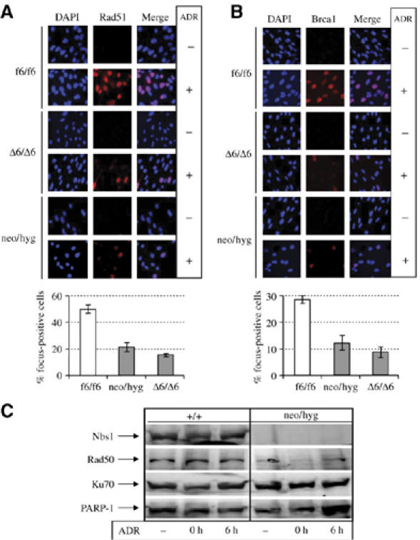
Defective focus formation of DSB repair molecules in MEF cells lacking Nbs1. Immunofluorescence analysis of Rad51 (A) and Brca1 (B) focus formation in inducible (Δ6/Δ6) and constitutive (neo/hyg) Nbs1-null MEFs after treatment with 0.2 μg/ml ADR for 3 h. Lower panels of (A) and (B) are quantifications of DNA damage-induced foci. Bars represent means±s.d. are shown for duplicate samples from two independent experiments. For each sample at least 500 nuclei were scored. (C) Western blot analysis of the chromatin-enriched fraction of MRN and Ku70 proteins. 0 h, 0 h after removal of ADR; 6 h, 6 h after removal of ADR. PARP-1 is served as a control.