
Figure 1.
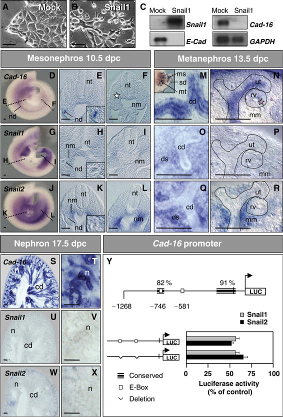
Snail1 represses the kidney epithelial Cadherin-16 both in cell culture and in the embryo. (A, B) Phase-contrast images and (C) Snail1, E-Cadherin and Cadherin-16 expression in stable mock- and Snail1-transfected cells. Snail1 expression represses E-cadherin and Cadherin-16 transcription. GAPDH levels are shown as a control. (D–X) ISH for Cadherin-16, Snail1 and Snail2 in whole-mount mouse embryos and transverse sections taken at the mid (E, H, K) and posterior (F, I, L) trunk levels. Cadherin-16 is expressed in the newly formed nephric duct epithelium (nd, E) that no longer expresses Snail genes (H, K insets). Snail transcripts are observed in the undifferentiated anterior (H, K) and posterior (I, L) nephrogenic mesenchyme (nm). Dissected urogenital system (see M, inset) or gelatin sections (M–R) hybridized with Cadherin-16 and Snail probes. Cadherin-16 is expressed in the collecting duct epithelia (M) and their ureteric tips (ut, N) of the developing metanephros but it is absent from the newly forming renal vesicle (pink star, rv; N). Expression is also detected in the sexual ducts and in the tubules of the transient mesonephros (sd and ms; M, inset). Snail1 and Snail2 expression is restricted to the metanephric mesenchyme and to the deep stroma (mm and ds, O–R). Nephrons (n) appear after the complete epithelialization of the metanephric mesenchyme. The nephron epithelia and the collecting ducts (cd) strongly express Cadherin-16 (S, T), whereas Snail1 and Snail2 expression disappears when the mesenchyme transforms into epithelia (U–X). nt, neural tube. Scale bar, 100 μm. (Y) Snail proteins repress the Cadherin-16 promoter. Schematic representation of the mouse Cadherin-16 promoter showing the regions of high similarity between mouse and human, and the location of the consensus Snail-binding sequences (white boxes). Luciferase reporter constructs carrying the wild-type mouse Cadherin-16 promoter (−1268) or deletions in the two E-boxes were assayed in the NMuMG cells together with either the mouse Snail1 or Snail2 expression vectors or an empty vector as a control (pcDNA3). Luciferase activity was measured 24 h after transfection and the activity is expressed relative to that of the wild-type construct. The results are the mean values±S.E. of duplicates from four independent experiments. Deletions of the Snail-binding sites do not relieve the repression of the Cadherin-16 promoter activity.