
Figure 3.
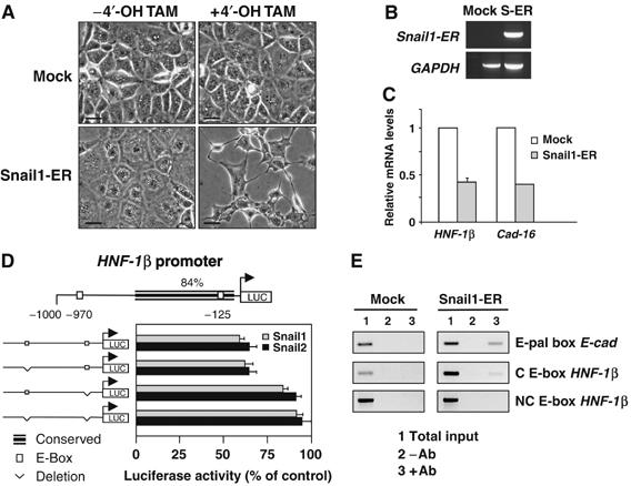
Snail genes directly repress HNF-1β transcription, which in turn impairs Cadherin-16 expression. (A) NMuMG cells stably transfected with an inducible Snail1 construct (Snail1-ER) or with the empty vector (Mock) were analyzed 24 h after induction. Note the phenotypic change of the Snail1-transfected cells upon 4′-OH-tamoxifen (4′-OH-TAM) administration. Scale bar, 25 μm. (B) Transgene expression visualized by RT–PCR in Mock and Snail1-ER transfectants (S-ER). (C) Quantitative RT–PCR for Cadherin-16 and HNF-1β 24 h after 4′-OH-tamoxifen administration. (D) Diagram of the 1 kb region upstream of the translational initiation site in the mouse HNF-1β gene showing regions highly conserved between mouse and human. Of the two consensus E-boxes for Snail binding, only one lies within the conserved region. Snail1 and Snail2 repressed the activity of the wild-type HNF-1β promoter (measured as described for the Cadherin-16 promoter), but they did not affect the promoter constructs in which the conserved E-box was deleted. (E) ChiP analyses show that Snail1 binds directly to the HNF-1β promoter. ChIP assays were carried out with anti-ER antibodies on Mock- and Snail1-ER cells 24 h after induction. As a positive control, the interaction of Snail with the E-pal element of the E-cadherin promoter is shown. Snail does not bind to the nonconserved (NC) E-box in the HNF-1β promoter. Amplifications of the indicated promoter regions in the input (1), nonimmunoprecipitated (2) and immunoprecipitated (3) fractions are shown. The data presented are representative of three independent experiments.