
Figure 6.
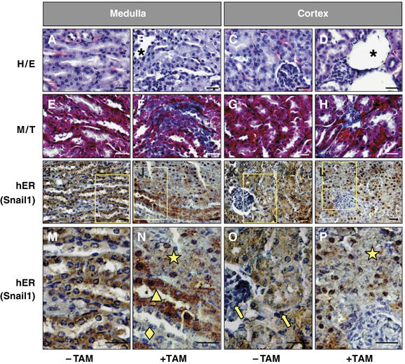
Snail activation is sufficient to induce renal fibrosis in adult transgenic mice. (A–D) Hematoxylin/eosin staining of sections from 4-month-old transgenic mice kidneys. Tamoxifen treatment in mice (+TAM) was initiated 2 months after birth. Note the defective overall morphology including the presence of dilated tubules. (E–H) Fibrotic deposits can be observed by Masson–Trichrome staining in tamoxifen-treated mice. Fibrosis is also manifested by the presence of cysts (D). (I–P) Detection of the transgenic Snail1 protein with an anti-hER antibody (brown) in paraffin sections counterstained with hematoxylin (blue). Note the absence of transgenic protein in the glomeruli and in the interstitial cells (yellow arrows in O) and in some ducts in the medulla (yellow diamond in N). The yellow triangle in (N) indicates ducts in which the Snail1 protein has not been efficiently translocated to the nucleus (note the blue nuclei and brown cytoplasms). The ducts and tubules with Snail1 nuclear expression lost the epithelial character and present a completely disorganized structure (yellow star in N and P). Scale bars, 25 μm.