Figure 7.

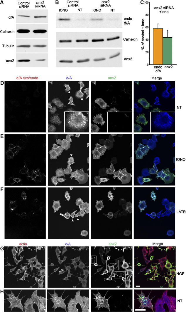
Downregulation of anx2 blocks enlargeosome exocytosis. (A) bottom row: compared to scrambled siRNA (control), anx2 siRNA induces in PC12-27 a considerable decrease of the protein without any parallel decrease of d/A, calnexin and tubulin. (B) Compares the amount of d/A mAb endocytosed by cells treated with either the scrambled or the anx2 siRNA, exposed (IONO) or not (NT) to ionomycin. The cells treated with anx2 siRNA, which express decreased levels of anx2 (bottom row) and normal levels of calnexin (middle row), show greatly reduced exo/endocytic responses. Quantification (averages of three experiments) of the endocytosed anti-d/A Ab and of anx2 in siRNA-treated cell populations exposed to ionomycin is shown in (C). The immunofluorescence of the d/A exo/endocytosis (exo/endo), and of anx2 and d/A expression in PC12-27 cells treated with anx2 siRNA, non-treated (NT) or stimulated with ionomycin (IONO), is shown in (D, E). Of the cells expressing d/A, only some are positive also for anx2 (merge image in the right frame), and only the latter show enlargeosome exo/endocytosis upon ionomycin, whereas the others show no appreciable responses (d/A exo/endo, left frame). (F) Analogous results obtained using latrunculinA (LATR), instead of ionomycin, to stimulate exocytosis. Actin (revealed by phalloidin), d/A and anx2 triple immunolabelling of a group of anx2 siRNA-treated PC12-27 cells exposed to NGF (2 days) is illustrated in (G) at higher magnification, and in non-differentiated cells, in (H). Only the cells maintaining normal expression of anx2 (squares in the anx2 frame) exhibit long dendrites, whereas those showing downregulation of the protein do not. Actin cytoskeleton maintains the usual morphology also in cells depleted of anx2. The bar in the merged frame of (G), valid also for (D–F), and that in the merged frame (H) correspond to 10 μm.
