
Figure 5.
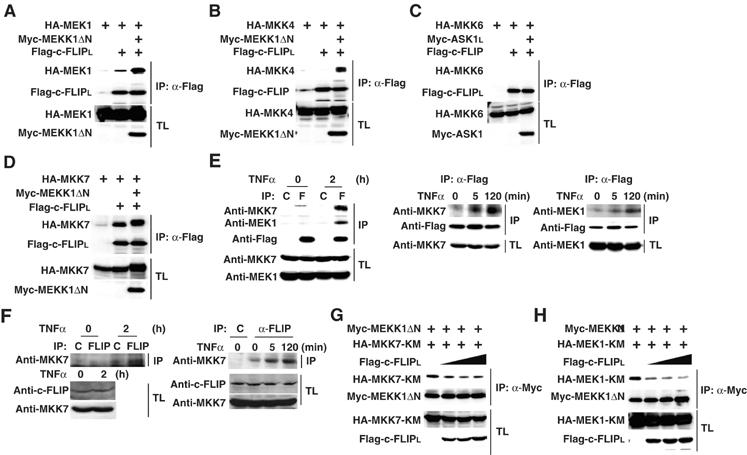
Interactions of c-FLIPL with MEK1 and MKK7 are induced in a TNFα-dependent, and c-FLIPL disrupts their interactions with MEKK1. (A–D) HEK293 cells were transfected with the indicated expression vectors. After immunoprecipitation (IP) with anti-Flag antibody, co-immunoprecipitated MAPKKs were detected by immunoblotting with anti-HA antibody (top panels). Expression levels of the transfected proteins in the immunoprecipitates (IP) (second panels) and the total lysates (TL) (third and bottom panels) were analyzed by immunoblotting with anti-Flag, anti-HA, and anti-Myc antibodies. (E) RelA−/− MEFs stably expressing Flag-c-FLIPL were stimulated with TNFα (10 ng/ml) for the indicated times. After immunoprecipitation with control (C) or anti-Flag (F) antibody, co-immunoprecipitated MKK7 and MEK1 were detected by immunoblotting with anti-MKK7 and anti-MEK1 antibodies, respectively. Expression levels of Flag-c-FLIPL in the immunoprecipitates (IP), MKK7 and MEK,1 in the total lysates (TL) were analyzed by immunoblotting with the indicated antibodies. (F) HEK293 cells were stimulated with TNFα (10 ng/ml) for the indicated times. After immunoprecipitation with control (C) or anti-c-FLIP antibodies, co-immunoprecipitated MKK7 was detected by immunoblotting with anti-MKK7 antibody (top panel). Expression levels of c-FLIPL and MKK7 in the total lysates (TL) (second and bottom panels) were analyzed by immunoblotting with the indicated antibodies. (G, H) HEK293 cells were transfected with the indicated expression vectors. After immunoprecipitation (IP) with anti-Myc antibody, co-immunoprecipitated MKK7-KM or MEK1-KM were detected by immunoblotting with anti-HA antibody (top panels). Expression levels of the transfected proteins were analyzed as in (A).