
Figure 2.
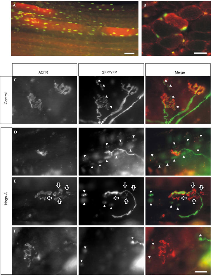
Overexpression of Nogo-A in muscle causes the disassembly of the neuromuscular junction. Electroporation of the soleus muscle with expression constructs for Nogo-A and nuclear-localized green fluorescent protein (GFP) results in muscle fibres that express high levels of Nogo-A (red) and GFP (green) after 2 weeks. (A) Whole-mount and (B) cross-section of the soleus muscle. Representative pictures of neuromuscular junctions (NMJs), 6 weeks after electroporation of empty vector (control; C) or Nogo-A expression constructs (D–F). The postsynaptic apparatus was visualized by rhodamine-labelled α-bungarotoxin (acetylcholine receptor; red in merge). Presynaptic nerve terminals were labelled by yellow fluorescent protein (YFP; green in merge) and transfected muscle fibres by nuclear localization signal–GFP (GFP, green in merge). For clarification, GFP-positive myonuclei are indicated by arrowheads. In control transfections, both pre- and postsynaptic structures look normal (C). By contrast, overexpression of Nogo-A often results in smaller NMJs (D) and fragmentation of the postsynaptic structure including partial (E; hollow arrows) or total (F; asterisks) retraction of the presynaptic nerve terminal. Scale bar, 25 μm.