
Figure 1.
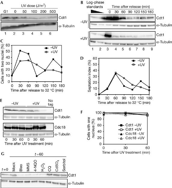
DNA damage reduces Cdt1 levels in M- and G1-phase cells. (A) Western blot analysis of Cdt1–Myc levels after ultraviolet (UV) irradiation of nitrogen-starved G1-arrested cells (P1424). Cells used for lanes 2–6 were released from the block for 30 min by re-feeding at 34°C. (B) Western blot of Cdt1–Myc levels in the mitotic cell cycle after UV irradiation. nda3 cells (P1451) were arrested in mitosis, UV irradiated (100 J/m2) and then released from the block by shifting to 32°C. Log-phase extracts are shown for comparison. (C) Nuclear counts for cells in experiment shown in (B) showing the timing of anaphase. (D) Septation index of cells for experiment shown in (B). (E) Western blot analysis of Cdt1–Myc and Cdc18–TAP levels in mitotically arrested cells after UV irradiation (100 J/m2). Strains P1451 and P1452 were treated as in (B), except that cells were kept at 20°C after irradiation to maintain the mitotic arrest. (F) Nuclear counts of the experiment shown in (E), showing that UV irradiation does not lead to the release of cells from the nda3 mitotic block. (G) Cdt1–Myc levels after treatment with DNA damage and stress agents. Cells (P1451) were arrested at the nda3 block, treated with 10 μg/ml bleomycin (bleo), 0.2% MMS, 0.6 μg/ml 4-NQO, 0.5 mM hydrogen peroxide (H2O2), 32 μg/ml chloroquin (CQ), 0.5 mM CdSO4 or 1 M sorbitol, and extracts were prepared 60 min later. Cells were kept at 20°C to maintain the mitotic arrest. MMS, methyl methane sulphonate; 4-NQO, 4-nitroquinoline 1-oxide; TAP, tandem affinity purification tag.