
Figure 3.
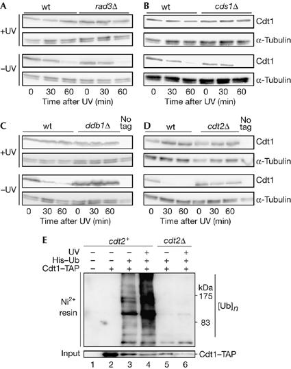
Ultraviolet-induced proteolysis of Cdt1 is not dependent on Rad3 and Cds1, but requires Ddb1 and Cdt2. (A) Cdt1–Myc levels after UV treatment in nda3 (wild type (wt), P1451) and nda3 rad3Δ cells (P1630) arrested at 20°C. (B) Cdt1–Myc levels after UV treatment in nda3 (wt, P1451) and nda3 cds1Δ (P1623) cells arrested at 20°C. (C) Cdt1–Myc levels after UV treatment in nda3 (wt, P1451) and nda3 ddb1Δ (P1615) cells arrested at 20°C. (D) Cdt1–Myc levels after UV treatment in nda3 (wt, P1451) and nda3 cdt2Δ (P1706) cells arrested at 20°C. In (A–D), the mitotic arrest was maintained throughout the time course and all UV treatments were carried out 100 J/m2. (E) High-molecular-weight ubiquitylated Cdt1 levels are increased after UV irradiation in cdt2+ strains. Ubiquitylated proteins were purified under denaturing conditions from strains expressing His6–ubiquitin (His-Ub) by Ni2+-NTA agarose affinity chromatography and probed with peroxidase–anti-peroxidase soluble complex to detect Cdt1–TAP. Irradiated strains were exposed to UV (100 J/m2) and grown for 20 min before preparing extracts. Strains used were P137 (lane 1), P1517 (lane 2), P1783 (lanes 3,4) and P1785 (lanes 5,6). NTA, nitrilo-triacetic acid; TAP, tandem affinity purification tag.