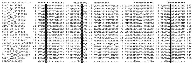
Figure 1.
Multiple sequence alignment of the RuvC family of HJ resolvases. The alignment was constructed by using the macaw program; only the five conserved motifs are shown. The lengths of the poorly conserved spacers between the motifs are indicated by italic numbers. The positions of the first and the last of the aligned amino acid residues in each sequence are also indicated. The protein designation consist of the gene, an abbreviated species name, and the GenBank identification number (separated by underlines). The consensus derived using 90% conservation is shown underneath the alignment; b indicates “big” residues (E, K, R, I, L, M, F, Y, W), h indicates hydrophobic residues (A, C, F, I, L, M, V, W, Y), s indicates small residues (A, C, S, T, D, N, V, G, P), u indicates “tiny” residues (G, A, S), and p indicates polar residues (D, E, H, K, N, Q, R, S, T). The conserved acidic residues constituting the catalytic triad of RuvC are highlighted by shading with reverse lettering. The multiple-alignment-based secondary structure prediction is shown on top of the alignment; E (e) indicates extended conformation (β-strand), and H (h) indicates α-helix (uppercase indicates the most confident prediction). Species abbreviations: Bacteria: Ct, Chlamydia trachomatis; Ec, E. coli; Hp, Helicobacter pylori; Mtu, Mycobacterium tuberculosis; Rp, Rickettsia prowazekii; Ssp, Synechocystis sp.; Tm, Thermotoga maritima; Tp, Treponema pallidum. Eukaryotic mitochondrial: Sc, Sac. cerevisiae; Sp, Sch. pombe. Viruses: biL66, LBPc2, Lactococcus lactis bacteriophages; CIV, Chilo iridescent virus; MCV, molluscum contagiosum virus; MSEPV, Melanoplus sanguinipes entomopoxvirus; Myx, myxoma virus; RFV, rabbit fibroma virus; VACC, vaccinia virus; YMV, Yaba monkey virus.

