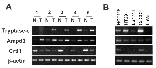
Figure 3.
RT-PCR analysis of human colon cancer tissue and cell lines. (A) RT-PCR analysis was performed on 5 paired normal and tumor human colon cancer tissues. T indicates tumor tissue and N indicates corresponding normal adjacent mucosa. Gene-specific primers for PCR were designed by MacVector 7 software depending on the information from GeneBank. Amplication of the right target DNA was confirmed by sequence analysis. β-actin was used as an internal control to confirm equal amount of the templates. (B) RT-PCR analysis was performed on 5 human colon cancer cell lines (HCT116, HT29, LS174T, CaCO2, LoVo) with the indicated primer sets.

