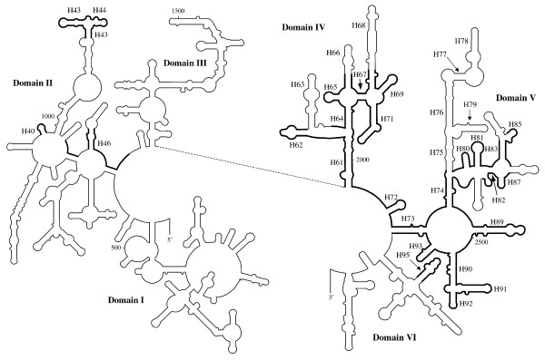
Figure 3.

Schematic diagram indicating the extent of the LSU rRNA gene in Amphidinium. Structural model of the folding of the 5' and 3' halves of LSU rRNA from E. coli. Regions where homologous sequence or structure is found on Amphidinium minicircles are in bold. The proposed base-pairing of the individual structures is displayed in additional file 11, along with detailed numbering of the positions of the structures. Helices proposed to be present in Amphidinium or that are mentioned in the text are labelled (e.g. H18 for helix 18). Helix numbering as defined by Ban et al.[26].
