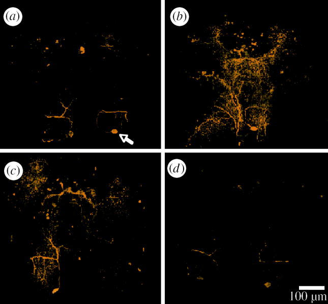
Figure 3.

Images show 5-HT immunoreactivity (yellow) within the brains of (a) uninfected (arrow shows position of TGN cell body) and (b) P. laevis-, (c) P. tereticollis- and (d) P. minutus-infected individuals. No differences, gross or fine, in brain anatomy from infected and uninfected individuals were observed. Bar shows 100 μm.
