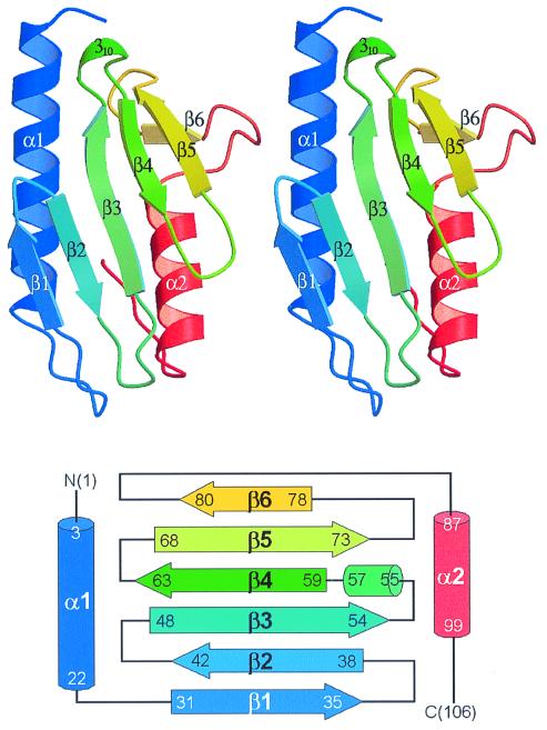
Figure 2.
Overall fold of E. coli CyaY. (Upper) Stereo ribbon diagram showing the secondary structure elements. Six β-strands (arrows), two α-helices (ribbons), and a 310-helix are drawn and labeled. molscript (23) and raster3d (24) programs were used to generate the figure. (Lower) Topology diagram.

