
Figure 3.
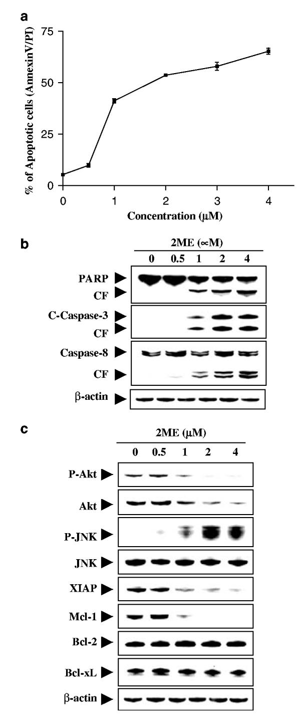
2ME markedly induces apoptosis in Jurkat acute T-cell leukemia cells in a dose-dependent manner. Jurkat cells were treated without or with various concentrations of 2ME as indicated for 24 h. (a) Cells were stained with annexin V/PI, and apoptosis was determined using flow cytometry as described in Materials and methods. The values obtained from annexin V/PI assays represent the mean±s.d. for three separate experiments. (b) Total cellular extracts were prepared and subjected to Western blot analysis using antibodies against PARP, cleaved caspase-3, and caspase-8. (c) Total cellular extracts were also prepared and subjected to Western blot assay using antibodies against phosphor-Akt, Akt, phospho-JNK, JNK, XIAP, and Mcl-1. For Western blot analysis, each lane was loaded with 30 μg of protein; blots were subsequently stripped and reprobed with antibody against β-actin to ensure equivalent loading. Two additional studies yielded equivalent results