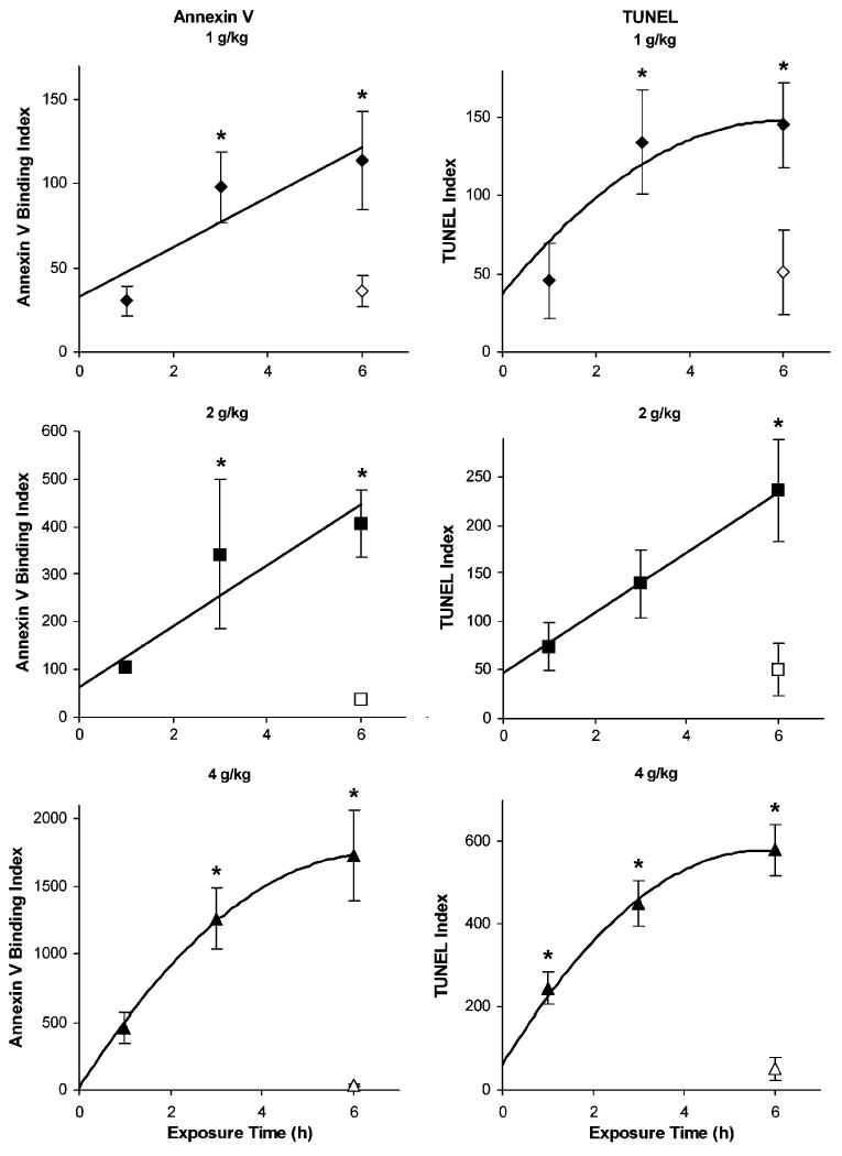
Fig. 3.

Time dependency and dose dependency of cell death in embryos exposed to ethanol in utero. Annexin V binding (left panels) and terminal deoxynucleotidyl transferase-mediated dUTP-X nick end labeling (TUNEL; right panels) were quantified in E7.5 embryos, as described in the Materials and Methods section, during the first 6 hours after treating pregnant females with 1 (diamonds), 2 (squares), or 4 (triangles) g/kg ethanol. Pregnant dams treated for 6 hours with maltose–dextran served as control embryos (open symbols). Mean and SE are shown. N = 59 to 10 embryos per treatment. *p<0.05 compared to control embryos.
