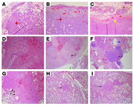
Figure 5. TβRIII decreased tumor cell invasiveness and metastasis in vivo.
Representative H&E staining (original magnification, ×10) of (A and B) primary tumors from mice implanted with 4T1-Neo cells exhibiting local invasion (red arrows) of tumor cells into the adjacent normal mammary tissue (A) and skin (B); (C) a representative primary tumor from mice implanted with 4T1-TβRIII cells demonstrating the absence of local invasion, as indicated by the clear margin between the tumor and the adjacent normal mammary tissue (yellow arrow); (D) a recurring tumor in a mouse at the primary injection site of 4T1-Neo cells exhibiting internal bleeding due to invasion of tumor cells into the blood vessels; (E) a metastatic tumor (black arrow) adjacent to the pancreas (green arrowhead) found on the mesentery of a mouse implanted with 4T1-Neo cells; (F) a significantly enlarged paratracheal lymph node adjacent to the trachea (blue arrowhead) containing metastatic tumor cells (black arrow) in a mouse with 4T1-Neo cells, indicating the presence of lymphatic metastasis; (G) multiple large metastatic tumor nodules (black arrows) in the lung of a mouse implanted with 4T1-Neo cells; and (H and I) representative lung metastases in mice implanted with 4T1-TβRIII cells (black arrows).

