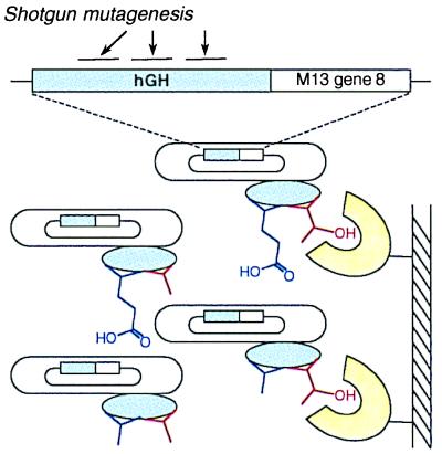
Figure 1.
Scheme for hGH shotgun scanning. The hGH gene was fused to M13 gene-8, and an hGH library was constructed with 19 mutated positions on three noncontiguous stretches of primary sequence. The library was then phage displayed, resulting in phage particles with hGH variants (blue ovals) displayed on their surface and the cognate hGH genes encapsulated within their coats. For simplicity, we show only two scanned side chains. Mutated positions initially had an approximately even distribution of wt and alanine. After selection for binding to an immobilized ligand (e.g., hGHbp shown in yellow), enrichment for wt side chains was observed for residues that contribute favorably to binding (red side chain). No selection for the wt amino acid was observed for residues that do not contribute to the binding interaction (blue side chain).

