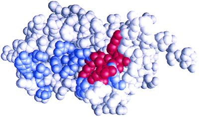
Figure 2.
Shotgun scan of hGH site 1 for binding to the hGHbp. The three-dimensional structure of hGH (23) is shown in space-filling mode. The 19 shotgun-scanned residues are colored, with blue denoting ΔΔGmut-wt < 1.0 kcal/mol and red denoting ΔΔGmut-wt > 1.0 kcal/mol (Table 2). The residues shown in red constitute the functional epitope of hGH site 1. The figure was produced with grasp software (30).

