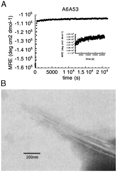
Figure 3.
(A) Seeding experiment followed by MRE at 217 nm for A6A53 variant at pH 5.2, 318 K, in 50 mM sodium acetate buffer. (Inset) The second phase of the seeding experiment, from 600 s to 21,600 s. (B) Electron micrograph showing the fibrils from auto-seeding experiment for A6A53 variant at pH 5.2 in 50 mM sodium acetate buffer.

