Abstract
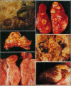
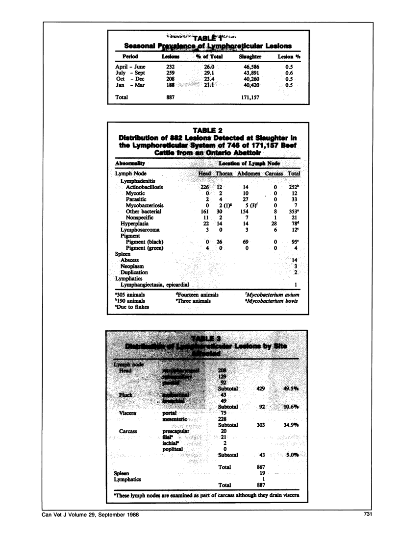
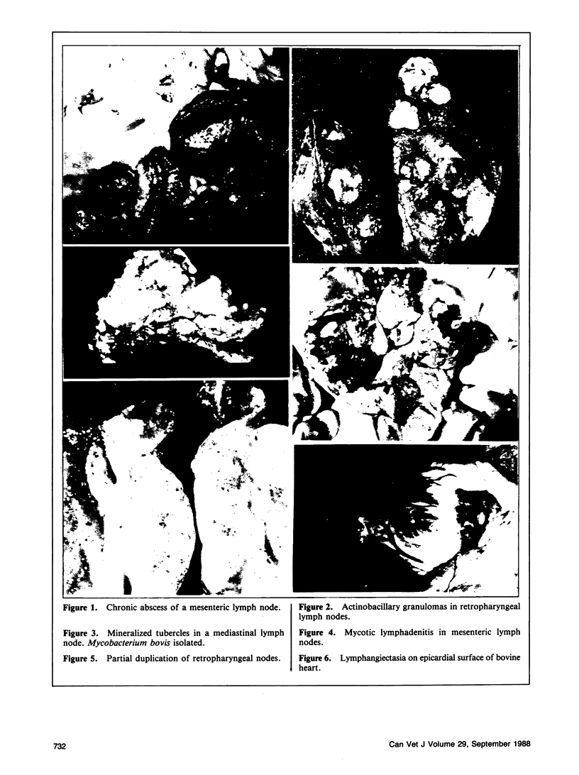
During the period April 1983 to March 1986, lymphoreticular lesions in cattle were surveyed at an Ontario abattoir. Postmortem examination of 171,157 cattle revealed macroscopic lesions in 696 animals (0.4%). The most frequent finding was abscessation of a single lymph node, a finding that was observed in 353 cases (50.7% of animals with lesions/0.2% of total slaughter). Actinobacillary granulomas were present in 252 lymph nodes (36.2%/0.1%). Other specific lesions included mycobacteriosis and mycotic or parasitic lymphadenitis. Cases of nonspecific chronic lymphadenitis or granulomas in lymph nodes, pigmentations, malformations, hyperplasia, and neoplasia were also seen. Abscesses were the most common splenic lesions. One animal had localized lymphangiectasia of the epicardium.
Full text
PDF
Images in this article
Selected References
These references are in PubMed. This may not be the complete list of references from this article.
- Angus K. W., Gilmour N. J., Dawson C. O. Alimentary mycotic lesions in cattle: a histological and cultural study. J Med Microbiol. 1973 May;6(2):207–213. doi: 10.1099/00222615-6-2-207. [DOI] [PubMed] [Google Scholar]
- Dion W. M., Bundza A., Dukes T. W. Mycotic lymphadenitis in cattle and Swine. Can Vet J. 1987 Jan;28(1-2):57–60. [PMC free article] [PubMed] [Google Scholar]
- Dukes T. W., Bundza A., Corner A. H. Bovine neoplasms encountered in Canadian slaughterhouses: a summary. Can Vet J. 1982 Jan;23(1):28–30. [PMC free article] [PubMed] [Google Scholar]


