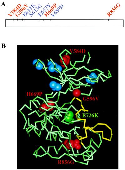
Figure 1.
(A) Point mutations in the KL domain of Tyk2 analyzed in this study. The KL domain is represented by the rectangle. Residues are color-coded as described below. (B) Predicted locations of Tyk2 KL residues on the structure of the IRK. IRK residues corresponding to the Tyk2 KL residues analyzed here are highlighted on the crystal structure of the inactive form of IRK (PDB code 1IRK), the activation loop of which is shown in yellow. Residues whose substitutions in Tyk2 had no functional consequences (E611K, S613G, E657V, Y689D) are in blue. Residues whose substitutions in Tyk2 led to loss of function are in red and are labeled. The residue corresponding to the HopT42 gain-of-function mutation (E726K) is in green.

