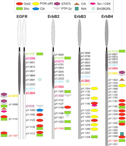
Figure 1.
Summary of systematic interaction profiling of the ErbB-receptor tyrosine family. All cytosolic residues of the ErbB-receptor family were used in pull-down assays. The interaction partners found are indicated by symbols. The kinase domain of the receptors is designated as an oval. Underlined and colored tyrosine residues mark identical sequence regions between different receptors; regions around colored residues show strong homology between receptors. Most interaction partners to tyrosine residues are found at the C-terminal end outside the kinase domain. The EGFR has multiple interaction partners, and several binding sites for Grb2. ErbB2 has few interaction partners; of them Shc is the most common. ErbB3 interacts mainly with P13-Kinase subunit p85, and ErbB4 again shows a diversity of interaction partners, also with several binding sites for Grb2. Receptors are not drawn to scale. The dimerization is indicated by EGFR dimer. Residues are labeled according to the full-length sequence of each of the receptors.

