Fig. 1.
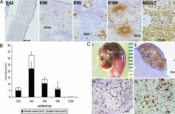
Histological markers of pig embryonic spleen before and after implantation. (A) Immunohistological staining of pig CD3 T cells in embryonic spleen tissue harvested at E42, E56, E80, E100, and adult spleen. (B) Morphometric analysis of implant growth. Different gestational ages were evaluated for total volume (white bars) and for vimentin volume-positive tissue (black bars) 6 weeks after transplantation (n = 5). (C) Development of E42 pig spleen graft under the kidney capsule of NOD-SCID mice. (Ci) Macroscopic view of E42 graft 6 weeks after transplant. (Cii) Pig mesenchymal components stained by anti-vimentin (V9). (Ciii) Porcine blood vessels stained by anti-pig CD31. (Civ) Proliferative status of the growing implant demonstrated by ki67 staining.

