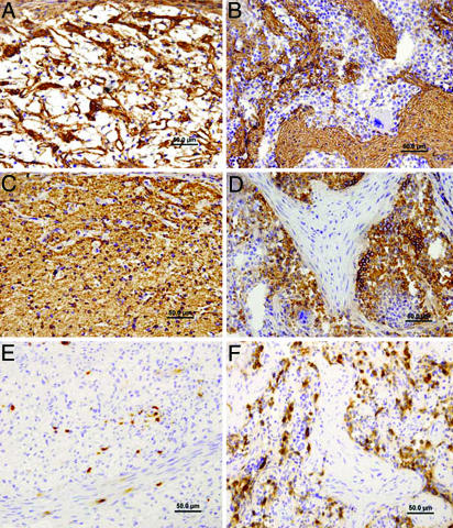
Fig. 2.
Development of hematopoietic nests and fibrous septae in pig E42 spleen implants. At 2 months, a sponge-like fibrous reticular network outlined by anti-laminin antibody (A) with diffusely entrapped mouse erythroid cells stained by anti-mouse TER-119 (C) is seen. In contrast, at 3 months after transplant, dense laminin-positive connective tissue septa is evident (B) surrounding nests of mouse hematopoietic tissue, including TER-119-positive erythropoietic areas, regions with megakaryocytes and myelopoiesis (D). Host myeloid cells, demonstrated by mouse Mac-1 immunostaining, are diffusely distributed in spleen transplants at 2 months (E) but become numerous within hematopoietic nests at 3 months after transplant (F).

