Fig. 3.
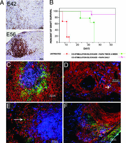
Engraftment of pig E42 embryonic spleen in Hu-SCID and C57BL/6 immunocompetent mice. (A) Rejection pattern of pig embryonic spleen mediated by Hu-PBMCs, 4 weeks after transplantation. Anti-human CD45 staining demonstrates reduced infiltration into grafts originating from E42 compared with E56 pig spleen. (B) Engraftment of E42 spleen under different immune suppression protocols. Engraftment pattern 7 weeks after transplantation in mice treated with rapamycin combined with costimulation blockade with anti-CD40L and CTLA4 is shown in C and E. Typically, the developing spleen tissue consists of periarteriolar lymphoid sheath with mouse T cells (CD3, blue) and a mouse B cell zone (CD45R, green). The pig stroma is denoted in red by staining with anti-vimentin. A few mouse macrophages (green) are shown on the periphery of the follicle of chimeric spleen (E, long arrow). In contrast, the rejection pattern at 2 weeks after transplantation, shown in D and F (in the absence of immune suppression), shows no evidence of follicle structures. Thus, mouse T cells (blue) and B cells (green, shown by short arrow) are diffusely spread on residual pig stroma (D), and numerous mouse macrophages (F4/80, green) (F) are detected in the implant.

