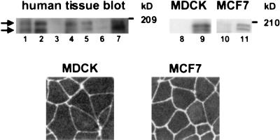
Figure 3.
(Upper) Distribution of AF-6 in multiple tissues and cell lines. (Lanes 1–7) A multiple human tissue blot from lung, kidney, spleen, testis, ovary, heart, and pancreas (Geno Technology, St. Louis) was immunoblotted with anti-AF-6 Ab. (Lanes 8–11) Immunoblot analysis of MDCK and MCF7 cell lysates with preimmune serum (lanes 8 and 10) and anti-AF-6 Ab (lanes 9 and 11), respectively. Two bands with molecular masses of approximately 195 and 180 kDa (kD) can be seen (arrows). (Lower) Localization of AF-6 in MDCK and MCF7 cells. Confluent MDCK and MCF7 cells were stained with rabbit polyclonal Ab against AF-6, followed by Texas red-conjugated anti-rabbit IgGs.

