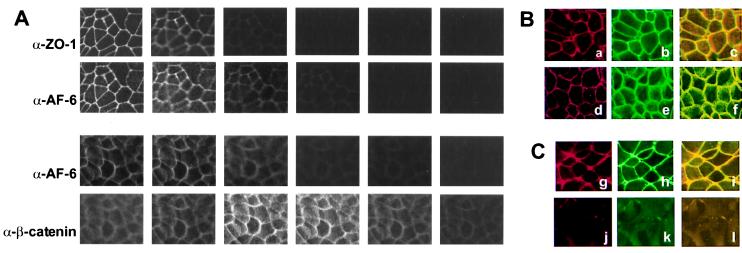
Figure 4.
(A) Confocal images of MCF7 cells comparing the localization of AF-6, ZO-1, and β-catenin. Confluent MCF7 cells were doubly stained with a rabbit polyclonal Ab against AF-6 and a mouse mAb against ZO-1 or a mouse mAb against β-catenin. As secondary Abs, Texas red-conjugated anti-rabbit IgGs and FITC-conjugated anti-mouse IgGs were used. Six serial optical sections (one section every 2 μm) are shown for each staining. From left to right, apical to basolateral side. (B) Localization of AF-6, ZO-1, and β-catenin in confluent control MCF7 cells. As described above, MCF7 cells were costained with a rabbit polyclonal Ab against AF-6 (a and d) and a mouse mAb against ZO-1 (b) or a mouse mAb against β-catenin (e). AF-6 is shown in red; ZO-1 and β-catenin are shown in green; and overlay is shown in yellow (c and f). (C) Localization of AF-6 and ZO-1 in Rap1E63-expressing (g–i) and Ha-RasV12-expressing (j–l) MDCK cells. The indicated cells were costained with a rabbit polyclonal Ab against AF-6 (g and j) and a mouse mAb against ZO-1 (h and k).

