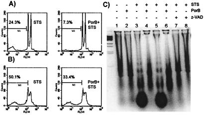
Figure 6.
Flow cytometry of PI fluorescence of Jurkat (A) and CH-12 RMC (B) cells incubated as described in the text. The percentage of cells containing hypodiploid DNA was measured by flow cytometry. The analysis gate has been drown accordingly to untreated samples (not shown). (C) DNA fragmentation pattern of CH-12 RMC cells incubated with: lane 1, medium alone; lane 2, 10 μg/ml PorB; lane 3, 1 μM STS for 4 h; lane 4, 10 μg/ml PorB for 24 h before STS incubation for 4 h; lane 5, 1 μM STS for 24 h; lane 6, 10 μg/ml PorB for 24 h before STS incubation for 24 h; lanes 7 and 8: 50 μM z-VAD-fmk before STS incubation for 4 and 24 h, respectively.

