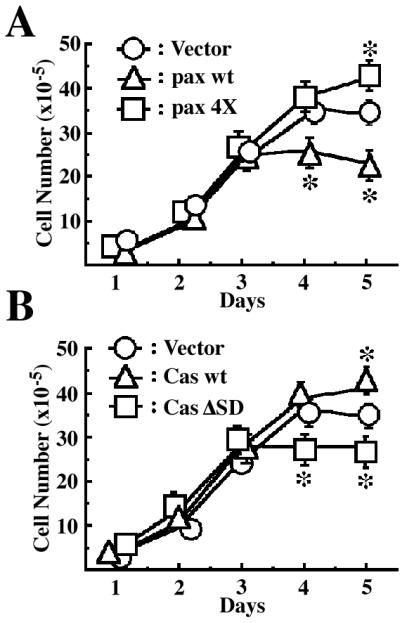
Figure 2.

Effects of EGFP-paxillin α, EGFP-p130Cas, and their tyrosine phosphorylation null mutants on cell saturation densities. Each 2 × 10⁵ cells were plated on 6-cm dishes and cultured for 5 days in the presence of serum. Cell numbers were measured every day by collecting cells and counting the viable cells. The same sets of NMuMG cells were used as in Fig. 1. Duplicate dishes were counted for each time point of the growth curves, and each determination represents the average of three independent experiments. Error bars represent the SEM. *, P < 0.05, as compared with the vector control cells.
