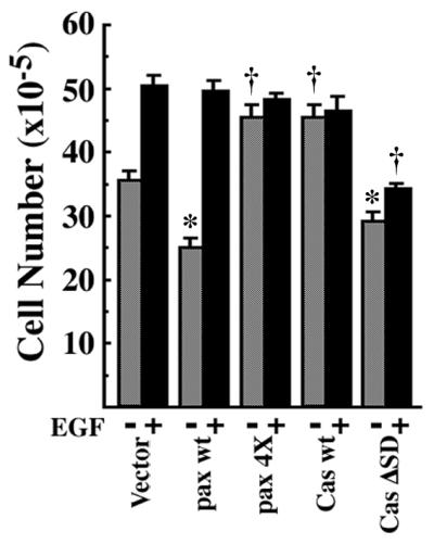
Figure 4.

EGF increases cell saturation density of NMuMG cells expressing paxillin α. The same sets of NMuMG cells were used as in Fig. 2. Each 2 × 10⁵ cells were plated on 6-cm dishes and cultured for 5 days in the presence of serum with (solid bar) or without (hatched bar) 10 nM EGF, and then viable cell numbers were counted. *, P < 0.05, †, P < 0.01 against values of the vector control.
