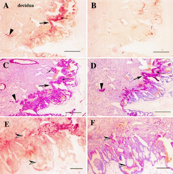
Figure 3.
Day 15 rhesus monkey implantation site. In situ hybridization for Mamu-AG mRNA. Serial paraffin sections of a day 15 implantation site were hybridized with Mamu-AG antisense (A and E) or sense probe (B), or stained with anticytokeratin mAb (C), anti-CD56 mAb (D), or anti-Sp1 Ab (F). Positive in situ hybridization signal or immunostaining appears red; cell nuclei are counterstained blue with hematoxylin. Location of the uterine decidua is noted in A in this and subsequent figures. (Scale bars: A–D, 500 μm; E and F, 100 μm.)

