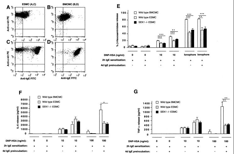
Figure 2.
ESMCs express functionally active FcɛRI receptors. (A–D) Flow cytometry analysis (104 cells/condition) of c-kit and FcɛRI expression in wild-type (129Sv) ESMCs (A and C) and BMCMCs (B and D) with (C and D) or without (A and B) preincubation with IgE (5 μg/ml) for 4 days (ref. 26). (E–G) Release of (E) β-hexosaminidase, (F) IL-6, or (G) TNF-α in wild-type BMCMCs, wild-type ESMCs, or SEK1 −/− ESMCs stimulated with IgE and specific Ag (DNP-HSA) (E–G) or calcium ionophore (E). Before stimulation, some cells (+) were preincubated with anti-DNP-HSA IgE (5 μg/ml) for 4 days to enhance FcɛRI expression. All cells were then sensitized with anti-DNP-HSA IgE (10 μg/ml) for 2 h and then stimulated with DNP-HSA or calcium ionophore. The percentage of of β-hexosaminidase release and cytokine release (23) were measured 1 or 6 h after Ag stimulation, respectively. In E–G, the results are mean ± SEM (n = 4–10) and are representative of the results obtained in two to three independent experiments. *, P < 0.05; **, P < 0.01; ***, P < 0.005, as compared with values for BMCMCs by unpaired Student's t test.

