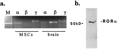
Figure 1.
Expression of RORα in hMSCs. (a) RT-PCR analysis using specific primers for RORα, RORβ, or RORγ as described in Experimental Procedures. RNA prepared from cells of different origin was used as a positive control (brain). M, size markers. (b) Immunodetection of RORα in hMSC nuclear extracts. Cell nuclei were prepared, and 40 μg aliquots of nuclei were separated on 10% SDS-polyacrylamide gel, transferred to plastic membranes, and probed with a polyclonal antibody raised against RORα.

