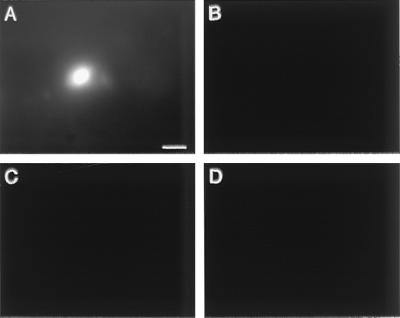
Figure 2.
Distribution of an adenoviral vector in tumor and liver after intratumoral injection. The control vector encoding the GFP protein but not an immunoconjugate was injected into three sites of a human melanoma skin tumor growing in SCID mice. The total vector dose was 6 × 10⁹ IU. The tumor and liver were dissected 40 h after the injection and were examined intact under a dissecting microscope with fluorescence optics. The GFP signal was detected with 480-nm excitation and 630-nm emission, and the background signal was detected with 577-nm excitation and 630-nm emission. (A) Tumor GFP. (B) Tumor background. (C) Liver GFP. (D) Liver background. A bright fluorescent spot similar to the one in A also was detected at two other tumor sites, presumably corresponding to the injection sites. The photographs are focused at one level in the tissues. However, the GFP spot in the tumor also could be detected by focusing above and below that level, suggesting that the tumor cells adjacent to the path traversed by the injection needle are the only cells infected by the vector. (Bar = 50 μm.)

