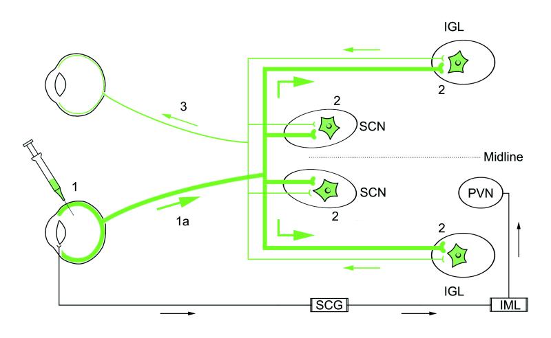
Figure 1.
A schematic diagram illustrating the neuronal circuits labeled after the intraocular injection of PRV 152. Primary infection of retinal ganglion cells occurs after intravitreal injection of PRV 152 (step 1). Viral replication and anterograde transport of virus (step 1a, thick green line) in axons of RGCs follows primary infection. Transsynaptic passage of virus from RGC axons produces secondary infection in a restricted set of retinorecipient central nuclei [i.e., SCN, IGL (shown), PT, and lateral terminal nucleus (not shown) as indicated by green (EGFP-labeled) neurons (2)]. Axon terminals of retinal ganglion cells in the contralateral retina, presynaptic to PRV 152-infected neurons in the SCN, IGL, PT, and lateral terminal nucleus, take up the virus via transsynaptic passage. The virus is retrogradely transported (thin green line) to the contralateral retina, where PRV 152-expressing ganglion cells are easily identified in whole-mount preparations (step 3). Intravitreal PRV 152 injection also infects autonomic afferents to the eye, resulting in retrograde transport of virus to the superior cervical ganglion (SCG), followed by transsynaptic uptake and retrograde transport to preganglionic neurons in the intermediolateral nucleus (IML) of the spinal cord (18). Neurons in the hypothalamic paraventricular nucleus (PVN) are subsequently infected after transsynaptic uptake of the virus from infected IML neurons and retrograde transport.

