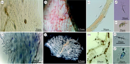
Figure 1.
Tissue and cells from extant ostrich and emu. (a–e) Emu. (a) Fibrous texture of demineralized cortical bone matrix. Osteocytes and vessel channels are visible. (b) Vessels emerging from demineralized cortical bone fragment, after partial enzyme digestion. Pigmented blood products fill vessel lumen. (c) Isolated vessel shows intimate association with osteocytes. (d,e) Isolated osteocytes with radiating filipodia, liberated after collagenase digestion of cortical bone. Variation in degree of pigmentation is seen, as well as variation in overall cell shape. (f–j) Ostrich. (f) Histochemical staining emphasizes fibrous texture of demineralized ostrich cortical bone matrix. Osteocytes are interspersed with collagen fibres. (g) Interconnecting lattice of vessels liberated from fresh ostrich cortical bone after demineralization and enzymatic digestion as above. (h) Ostrich vessel and associated matrix with osteocytes after demineralization and digestion. Blood breakdown products fill vessel lumen. (i) Unstained, isolated osteocyte is elongate with extensive filipodia. (j) Cell body is expanded and has been stained to reveal cellular detail. All specimens derived from 14+ year post-mortem specimen except (g), from ca 1 year post-mortem. Scale bars are as indicated.

