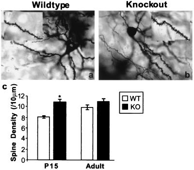
Figure 3.
Increased number of dendritic spines in young spinophilin knockout mice. (a and b) Representative Golgi staining of a neuron in caudatoputamen from wild-type (a) and knockout (b) littermates. (Bar = 100 μm.) (c) Spine density in wild-type (WT) and knockout (KO) littermates at P15 and adulthood. *, P = 0.01, compared with WT at P15.

