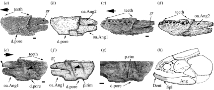
Figure 2.
Eoactinistia foreyi, NMV 218301, dentary in (a, b) lateral, (c, d) dorsal and (e, f) medial or internal views, (g) close up view of dentary pore, internal dentary surface, (h) Gavinia syntrips, representing a phylogenetically basal coelacanth (Middle Devonian, Australia), to illustrate the relationship between the dentary (grey) and other bones of the jaw. Note absence of dentary pore (from Long 1999). Scale, 200 μm; Ang, angular bone; d.pore, dentary pore; Dent, dentary bone; gr, narrow groove; oa.Ang1, 2, overlap surfaces for angular; p.rim, rim surrounding dentary pore; Spl, splenial bone; teeth, teeth associated with dentary in marginal tooth row. Larger arrows indicate anterior. ((h) Courtesy of Western Australian museum.)

