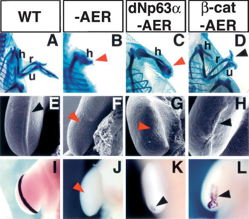
Figure 4.
β-Catenin induces AER and limb regeneration in chick embryos. (A–D) Cartilage staining of the chick forelimb at 10 d post-fertilization. (A) Wild-type chick limb showing the stylopod (humerus), zeugopod (radius and ulna), and autopod elements. (B,C) Removal of the AER at stage 20/21 prevented limb outgrowth and resulted in a truncated limb independent of dNp63α overexpression. (D) Ad-CA-β-catenin injection prior to AER removal restored limb development and induced the formation of all the stylopod, zeugopod elements, and two digits (black arrowhead). Red arrowheads in B and C indicate truncated limbs. (E–L) SEM images (E–H) and fgf8 expression (I–L) of the limb ectoderm 30 h after AER removal. The control limb shows the typical thickened AER ectoderm (black arrowhead, E) associated with fgf8 expression (I). Absence of distal thickened ectoderm after AER removal (red arrowhead, F) results in the down-regulation of fgf8 expression (red arrowhead, J). Overexpression of dNp63α did not rescue AER (red arrowhead, G) or fgf8 expression (residual expression is observed at the posterior of the developing limb bud) (black arrowhead, K). Infection of limb ectoderm cells with Ad-CA-β-catenin prior to AER removal regenerated the AER (black arrowhead, H) and elicited fgf8 expression (black arrowhead, L). (h) Humerus; (r) radius; (u) ulna.

