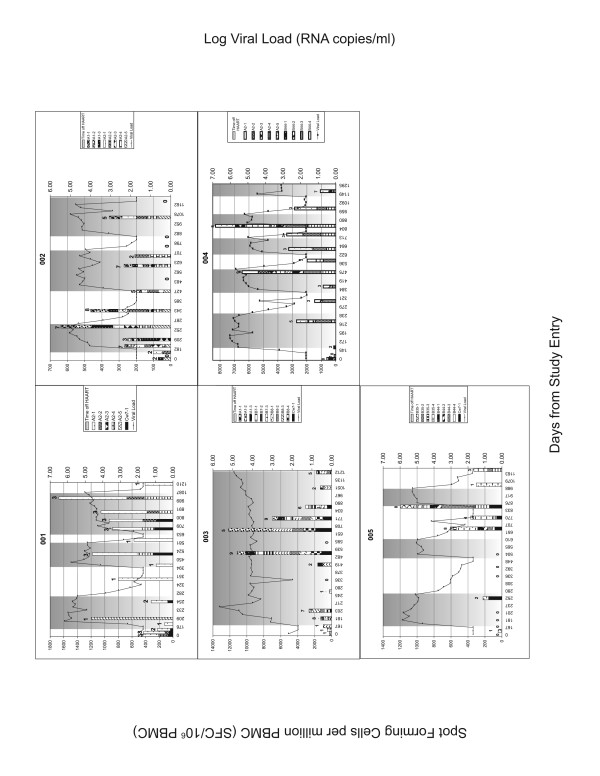
Figure 1.
Results of IFN-γ ELISPOT assay for patient 001 to 005. The left y-axis shows the number of spot forming cells (SFC)/106 PBMC. Each stacked bar shows the number of SFC/106 PBMC generated to the peptide panel tested at each clinic visit. The height of the stacks in each the bar represents the number of SFC/106 PBMC induced by each positive stimulus. The height of the bar is the cumulative magnitude of the response to the peptide panel tested. The number over the bar is the number of peptides in the panel recognized at that time point. The shaded areas are the intervals off HAART. Also shown are viral load determinations at each time point keyed to the right y-axis.

