
Figure 1.
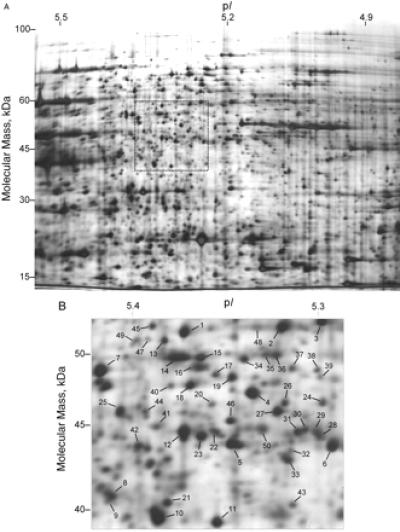
(A) Narrow pH range isoelectric focusing 2D gel (pH 4.9–5.5). Soluble yeast protein (500 μg) was loaded onto the gel. More than 1,500 features were visible by silver staining. (B) An arbitrary 4-cm2 region of the gel was selected for analysis. Numbers show the 50 spots that were identified by the MS techniques described in the text.