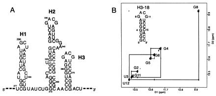
Figure 1.

Characterization of H3 stem loop sequence. (A) The secondary structure of the H1, H2, and H3 stem loops of Moloney murine leukemia viral RNA (10). (B) The sequence and secondary structure of H3–18; note that the two terminal G⋅C base pairs are different from the native sequence. The imino proton region of the water NOESY spectrum (mixing time 300 ms, 283 K, 100 mM NaCl, pH 6.5) of H3–18 confirms the base pairing. The G8 and G11 imino protons were assigned based on the evidence described in the text.
