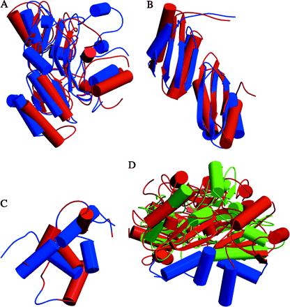
Figure 2.

Comparison of the three individual domains of d-LDH with the corresponding domains of PCMH and MurB. (A) The FAD-binding domains of d-LDH (blue) and PCMH (red). (B) The cap domains of d-LDH (blue) and PCMH (red). (C) The membrane-binding domain of d-LDH and the corresponding region in PCMH. (D) The overall structure of d-LDH (FAD and cap domains in red and the membrane-binding domain in blue) compared with MurB (green). The ribbon representation shows the general similarity of the FAD and cap domains of d-LDH to those of MurB or PCMH. The membrane-binding domain is absent in MurB and differs significantly in PCMH.
