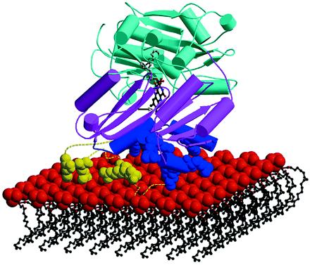
Figure 4.

Cartoon of d-LDH associating with the membrane. d-LDH is anchored to the membrane by electrostatic interactions between basic residues (blue balls) from the observed membrane-binding domain (blue) and possibly from the modeled missing segment (dashed yellow) comprising nine basic residues (yellow balls) and the negatively charged phospholipid head groups (red balls) of the membrane. The cap domain (pink) and FAD-binding domain (cyan) are also shown. The stick drawing of the FAD cofactor is depicted with gray balls for atoms in the adenine and sugar rings, with red balls for phosphate and oxygen atoms, and with black balls for atoms in the isoalloxazine ring. In this model, the substrate d-lactate can approach the active site (as shown by the black arrow) near the isoalloxazine ring (visible between pink strands) from behind the cap domain.
