
Figure 5.
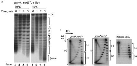
Topo IV efficiently relaxes positively supercoiled DNA in vivo. (A) pBR322 DNA was isolated from an E. coli ΔacrAparE10 strain after addition of 5 μg/ml novobiocin (Nov). Novobiocin was added at time 0, and cultures were either kept at 30°C (lanes 1–4) or shifted to 42°C (lanes 5–8). DNA was resolved on a 1% TAE gel with 10 μg/ml chloroquine. The positions of (−) supercoiled, [(−) sc], and (+) supercoiled, [(+) sc] topoisomers are shown. (B) pBR322 DNA was isolated from parC+ (Left) and parCr norfloxacin-resistant (Right) E. coli strains that had been treated with 100 μg/ml norfloxacin. The DNA was analyzed by two-dimensional agarose gel electrophoresis. The first dimension (1D) was a 1% TAE agarose gel without chloroquine, and the second dimension (2D) contained 10 μg/ml chloroquine. Reference plasmid DNA relaxed in vitro by calf thymus topoisomerase I is shown (Lower). The positions of (+) and (−) topoisomers are shown.