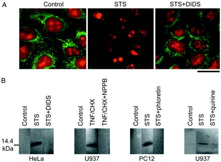
Figure 2.

Release of cytochrome c induced by treatment with an apoptogenic inducer, and its prevention by simultaneous treatment with a Cl− or K+ channel blocker. (A) Cytochrome c release from mitochondria monitored by immunocytochemistry in HeLa cells 5.5 h after treatment with STS in the absence or presence of 0.5 mM DIDS. A green fluorescence: mitochondrial cytochrome c revealed by FITC-conjugated secondary antibody. A red fluorescence: counter staining with propidium iodide. Data represent triplicate experiments. (Scale: 20 μm). (B) Cytochrome c release to cytosol was monitored by Western blot in U937, HeLa, and PC12 cells 8 h after treatment with STS or TNF/CHX in the absence or presence of 0.5 mM DIDS, 0.5 mM NPPB, 30 μM phloretin, or 0.5 mM quinine. Data represent triplicate experiments.
