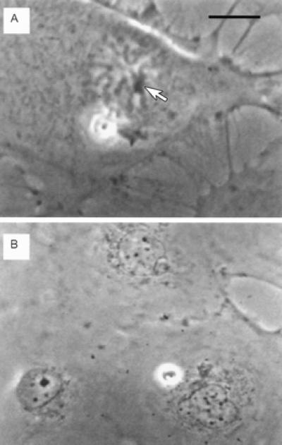
Figure 2.

Live phase contrast images of pre- and post-irradiation single nucleolar cell treated with ethidium monoazide bromide. (A) Pre-irradiation mid-prophase cell. One phase dense nucleolus can be seen clearly associated with condensed chromosomes (arrow). The nucleolus plus the closely associated chromosomes were exposed to the laser irradiation to ensure exposure of all of the ribosomal DNA. (Bar = 10 μm.) (B) The two daughter cells 24 h after laser exposure. Neither daughter cell has a nucleolus. Both have small micronuclear bodies that are typically found scattered throughout the nucleus. Note the size of the nucleolus and the three small nuclear bodies in the adjacent normal cell in this field. In earlier experiments (see ref. 9), inactivation of all of the ribosomal DNA by single-photon methods produced daughter cells with small nuclear bodies identical to the ones observed here. (Bar in A = 15 μm in B.)
