
Figure 3.
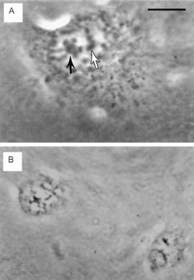
Live phase contrast images of pre- and post-irradiation double nucleolar cell treated with ethidium monoazide bromide. (A) Prophase cell with partially condensed chromosomes associated with two nucleoli (arrows). This cell was followed from the initiation of prophase to track the two nucleoli during the process of chromosome condensation. One nucleolus and its associated chromosome regions were irradiated (black arrow). (Bar = 10 μm.) (B) Two daughter cells 8 h after completion of mitosis. Both cells have only one nucleolus. The partially condensed chromatin visible in both nuclei generally disappears by 12 h after mitosis, when the cell enters the S phase of the cell cycle. (Bar in A = 15 μm in B.)