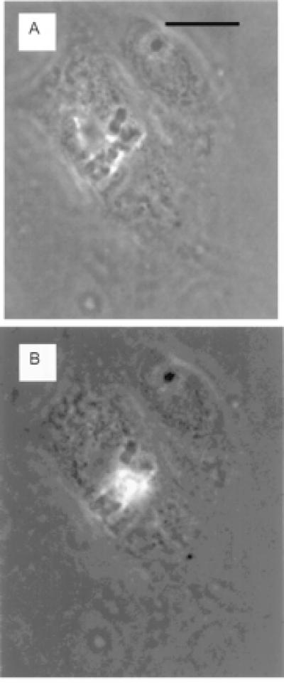
Figure 4.

(A) A mitotic cell with chromosomes clearly visible aligned at the metaphase plate. (B) The same cell with the chromosomes in the center of the metaphase plate exhibiting two-photon fluorescence during exposure to the 1.06-μm-wavelength laser beam focused through the ×63 microscope objective. Cells were exposed to ethidium monoazide bromide at 10 μm/1 ml for 24 h. (Bar = 15 microns.)
